
The healthy adult human brain consists of around 200 billion neuronal and non-neuronal cells that work together seamlessly. However, these cells contribute to brain function as discrete entities whose specific roles are still being determined; this information is vital to understanding the brain in health and disease. To fill the critical gap in knowledge about cell types that make up the human brain, the Brain Research Through Advancing Innovative Neurotechnologies® (BRAIN) Initiative has brought to bear collaborative and accelerated technology development to build tools for creating catalogs of cell types of the nervous system. Akin to cartographers, scientists are now developing detailed cellular maps of the brain in humans and other animals in order to better understand brain function.
Using high throughput technologies, cells that make up the brain are being catalogued and categorized at an unprecedented pace based on their molecular signatures and other physiological properties. Like coordinates on a map, the molecular identity of a cell offers the possibility of precision targeting for treating diseases. As catalogues of cells become richer with information, and more technological advances expand the toolkit for neuroscientists, BRAIN’s disease-agnostic investments in tool development are also fueling a better understanding of the cellular basis of brain dysfunction, ultimately paving the way for a new generation of precision therapeutics.
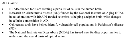
National Institute on Aging (NIA) funded study leverages BRAIN cell census tools for mapping the cellular changes in Alzheimer’s disease
Alzheimer’s disease (AD), a fatal disease marked by progressively worsening memory loss and dementia, is characterized by significantly atrophied brains. Determining which cells are lost in the brain and how this loss contributes to disease progression has presented a substantial challenge in the search for treatments for AD. However, a collaborative effort, which builds on research and tools developed under the aegis of the NIH BRAIN Initiative’s Cell Census Network (BICCN), is now mapping the cellular landscape of alterations in brain cell types in people with AD. A team of scientists at the Allen Institute, University of Washington School of Medicine and Kaiser Permanente Washington Health Research Institute, known as the Seattle Alzheimer’s Disease Brain Cell Atlas consortium, or SEA-AD, has released a large dataset of the molecular composition of 1.2 million brain cells from post-mortem brains donated by 84 people, comprising both healthy individuals and those with AD.
Funded by the NIA, detailed neuropathology as well as demographic, clinical and cognitive data for patients have also been released along with cellular data. A preliminary analysis suggests that the parts list of cells from the Alzheimer’s brain may differ significantly from the healthy brain. For example, inhibitory neurons, which reduce the activity of other neurons, show a substantial reduction in numbers, while some types of non-neuronal cell types increase in abundance relative to the healthy brain. These insights, along with other discoveries yet to come from this effort, packaged in publicly available datasets for clinicians to use, enable a technological shift in the search for the causes of AD. In return, a clear understanding of the make-up of the healthy brain in comparison to a brain afflicted with AD offers pathways to the development of better therapies.
A high-resolution map of cells suggests cell-autonomous dysregulation of specific genes in vulnerable cell populations in Parkinson’s Disease (PD)
Like AD, Parkinson’s disease (PD) is a chronic and progressive neurological disease. PD presents a significant and increasing threat to public health, with an estimated 1 million people who may have PD in the United States alone. A primary hallmark of the disease is the progressive loss of a class of neurons that release the neurotransmitter dopamine, located in a brain region called the substantia nigra. However, the substantia nigra itself consists of a variety of different classes of neurons, and identifying which ones go awry in PD raises the possibility of more effective treatments and long-term strategies to prevent the disease.
Work funded by the NIH BRAIN Initiative has now revealed with exquisite precision the human brain cell types that first stop functioning in Parkinson’s disease. By applying techniques to investigate the molecular composition of dopamine-releasing neurons in the substantia nigra in postmortem human brain tissue, scientists identified 10 distinct populations of neurons. Like assigning zip codes in a map, the researchers went on to map these cells to spatial locations within the substantia nigra. This combination of genomic techniques and spatial mapping allowed the scientists to identify a specific population of neurons, called SOX6_AGTR1 neurons and marked by the gene AGTR1, as being specifically lost in people with PD.
The same group of researchers went on to discover that SOX6_AGTR1 neurons carry within them several active genes previously implicated in genome-wide association studies of people with PD. Establishing the link between candidate genes for PD and the disease-susceptible neurons is remarkable because it suggests a cell-autonomous mechanism where specific genes within vulnerable SOX6_AGTR1 neurons malfunction. Researchers are now armed with a molecular handle to zero in on exactly how this might happen. Ultimately, understanding the molecular roots of Parkinson’s disease offers the best route to developing treatments and cures and perhaps even ways to prevent the disease entirely.
New funding opportunities at the National Institute on Drug Abuse (NIDA) applying cell census tools to study the effects of addictive substances in the brain
Genomic profiling of brain cells is also creating novel ways to investigate how substances such as opioids affect brain circuits, and the trade-off between their therapeutic benefits and addictive properties. Approximately 5.6 million Americans have an opioid use disorder (OUD), and approximately 8.7 million people misuse prescription opioids for durations, doses, or reasons other than those for which they were prescribed. The use of other classes of drugs, such as psychostimulants, has become more widespread among Americans, which has resulted in devastating increases in overdose deaths.
Brain circuits that elicit the feeling of reward and motivation in response to biological functions such as eating, finding mates, and learning new skills are central to the human experience. However, these same circuits for reward are vulnerable to addictive substances, and their dysregulation is thought to make some people susceptible to addiction. How brain circuits are co-opted and modified in response to substance use disorder or due to exposure to substances during sensitive periods of brain development are critical areas of research. Understanding how the brain changes in these circumstances may help address the lifelong impact and challenges posed by addiction and overdose. Now, cell census tools promise to provide new molecular insights that could be ultimately leveraged for treatments tailored to individual vulnerabilities.
The neural circuits that underlie the biology of reward and addiction are the subject of much active research, such as through the SCORCH (Single Cell Opioid Response in the Context of HIV) program, including the application of BRAIN-funded cell census tools to investigate opioid addiction. Two new funding opportunities at NIDA invite applications to use single-cell genomics tools such as those developed in the NIH BRAIN Initiative to investigate the effect of addictive substances on brain development at the cellular level (RFA-DA-23-036), and to map cell ensembles and cell types involved in opioid action in the rodent brain (RFA-DA-23-035):
-
RFA-DA-23-036: Investigating the effects of addictive substances on brain developmental trajectories using innovative scalable methods for quantification of cell identity, lineage and connectivity (R01-Clinical Trial Not Allowed)
-
RFA-DA-23-035: Large-scale integrated mapping and molecular profiling of cell ensembles and/or cell-types mediating opioid action in the rodent brain (R01-Clinical Trial Not Allowed)
The NIH BRAIN Initiative was launched with the audacious goal of revolutionizing our understanding of the human brain. Embedded in that goal is a promise that a better understanding of the human brain will pave the way for better treatments and cures of brain diseases and dysfunctions. Cell census tools developed with funding from the NIH BRAIN Initiative are now beginning to help chart that promise from basic research to applications for improving human health. Clinician-scientists can now begin to leverage new insights into the cellular basis of AD and PD – and the discoveries yet to come on the cellular changes in substance use disorders and other mental illnesses – to engineer new and better therapies for these devastating human brain disorders.